
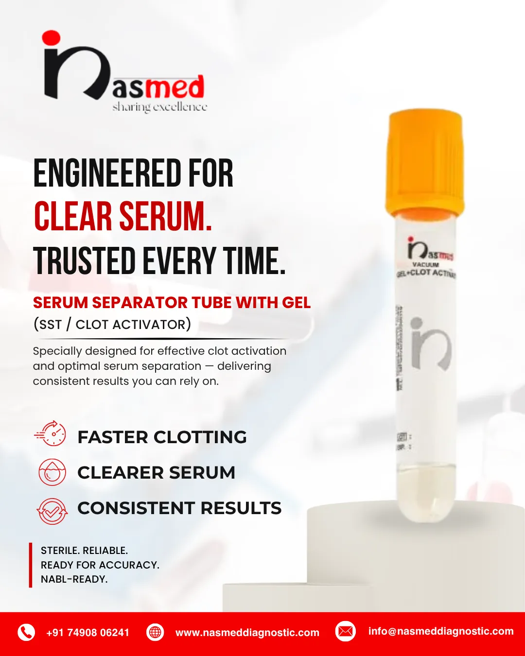
Serum Separator Tube (SST): Full Form, Uses, Tests and How It Works
SST stands for serum separator tube (also written serum-separating tube, and often called an SST tube, SST vial or gold-top tube). It is a vacuum blood collection tube that contains a clot activator and a separator gel. Once the blood has clotted and the tube is spun in a centrifuge, the gel forms a barrier between the clot and the serum above it, so the lab can run chemistry, hormone and serology tests on clean serum.
This guide explains what an SST is used for, which tests suit it, how the gel works, what the cap colours mean, and how to clot, spin and handle the tube correctly. If you work in a lab, a hospital or phlebotomy, it should help you avoid rejected samples and redraws.
SST at a glance
- Full form: SST = Serum Separator Tube
- Contains: clot activator (usually micronised silica) + inert polymer separator gel
- Cap colour: usually gold or red-and-grey marbled (“tiger top”); always confirm on the manufacturer’s label
- Sample produced: serum
- Typical use: routine chemistry, lipids, liver and kidney tests, thyroid and other hormones, cardiac markers, serology
What Is an SST? Full Form and Meaning
An SST is a sterile, vacuum-sealed tube designed to produce serum, the clear, straw-coloured liquid left after blood clots and the cells are removed. Most clinical chemistry tests run on serum.
Inside the tube you will find two key components:
- A clot activator, usually micronised silica, which speeds up clotting.
- A polymer separator gel, which sits at the bottom of the tube and forms a barrier after centrifugation.
Other Names and Cap Colours
People also call it a serum separator tube, serum-separating tube, SST tube, SST vial, serum gel tube, gold-top tube or tiger-top tube. The cap is typically gold or red-and-grey marbled, but colour coding can vary between manufacturers, so always check the label and the instructions for use.
Why the SST Matters Clinically
A doctor’s decision often rests on a single lab value. If cells stay in contact with serum, glucose falls and potassium rises, and the result misleads. The gel barrier keeps serum and cells apart, so the sample stays closer to the patient’s true condition. For a wider look at tube families, see this overview of types of blood collection tubes.
How Does a Serum Separator Tube Work?
The process has three stages: clotting, spinning and separating.
- Clotting. Blood enters the tube and touches the clot activator. Silica particles trigger the clotting cascade through contact activation of factor XII. Labs commonly allow about 30 to 60 minutes at room temperature, depending on the manufacturer’s instructions.
- Centrifugation. The clotted tube goes into a centrifuge. A commonly quoted setting is 1,000 to 1,300 RCF (relative centrifugal force, “g”) for about 10 minutes. Some laboratory manuals specify longer spins, for example 15 to 20 minutes depending on whether the rotor is swing-bucket or fixed-angle, so follow the tube insert and your lab’s procedure.
- Separation. During the spin the gel becomes thinner under force. Its density sits between that of blood cells and serum, so it moves up and settles between them. When the spin stops, it thickens again and holds its position.
What an SST Looks Like After Centrifugation
A properly spun tube shows three clear layers:Serum (clear, straw-coloured)Gel barrierClot (blood cells and fibrin)Illustration: layers in an SST after centrifugation (not to scale).
| Layer (top to bottom) | What it contains |
|---|---|
| Top | Clear serum, ready for analysis |
| Middle | Gel barrier |
| Bottom | Clot with blood cells |
If red cells or fibrin sit above the gel, or the gel barrier is incomplete, see the troubleshooting table further down.
What Is the Gel Inside an SST?
The separator gel is an inert, thixotropic polymer. “Thixotropic” simply means it flows when stirred or spun and firms up when left still. The exact chemistry is proprietary and varies by manufacturer, so the gel is usually described by its behaviour and density rather than a single universal name.
Why Gel Density Matters
Manufacturers formulate the gel to be heavier than serum but lighter than clotted cells. That balance lets it rise to the boundary between the two layers. If the density is off, the barrier forms poorly and serum can mix with cells again.
What the Gel Should Not Do
Good gel does not react with common analytes and does not release particles into serum. Still, some substances, such as certain therapeutic drugs and some hormones, can partly absorb into the gel. That is why some tests need a plain tube instead.
Are There “Glass Particles” in a Serum Separator Tube?
You may see this phrase in exam questions and textbooks. It refers to the clot activator: fine silica particles. Silica is the main component of glass, which is why the activator is sometimes described as “glass particles”. These particles activate clotting; they are part of the tube’s design, not contamination. Visible debris, cloudiness or foreign material in a sample is a different matter and should be reported to the manufacturer as a quality issue.
Clot Activator Options
Most SSTs use silica particles. Some “rapid serum” variants use thrombin instead, which clots blood faster. This helps in urgent settings or for patients on anticoagulant therapy.
Serum Separator Tube Tests: What Is an SST Used For?
Labs favour SSTs for routine chemistry because the serum is ready for automated analysers after one spin. Typical tests include:
| Test category | Examples |
|---|---|
| Metabolic panels | Basic and comprehensive metabolic panel (BMP, CMP), electrolytes |
| Lipid studies | Total cholesterol, HDL, LDL, triglycerides |
| Liver function | ALT, AST, ALP, bilirubin |
| Kidney function | Urea, creatinine |
| Thyroid and hormones | Thyroid profile and many other hormone assays |
| Cardiac markers | Cardiac enzymes and related markers |
| Serology | Hepatitis markers and many other immunology and infectious disease assays |
| Other serum tests | Iron studies, vitamin B12, vitamin D, folate, tumour markers (depending on your lab’s menu) |
The exact list differs from lab to lab. Your laboratory’s test directory always has the final word.
Tests That May Need a Different Tube
Some assays are sensitive to gel. Therapeutic drug monitoring, toxicology screens and certain specialised hormone tests often call for a plain or non-gel tube. Choosing correctly matters because a wrong tube can force a redraw. This practical reference on which tube to use for which test helps match tests to tubes.
SST Cap Colour: Gold, Tiger Top and Red-Grey
| Common name | Typical cap | Note |
|---|---|---|
| Gold-top SST | Gold or yellow | Most widely used name for a gel and clot activator tube |
| Tiger-top SST | Red-and-grey (or red-and-yellow) marbled | Same function: serum separator tube with clot activator |
| Red-top serum tube | Red | Usually no gel; not an SST |
Colour coding is a convention, not a guarantee. Manufacturers differ, so confirm the additive on the tube label before use.
SST vs Red Top vs PST (Plasma Separator Tube)
An SST and a red top both yield serum, but they get there differently. A PST (plasma separator tube, often light green) uses an anticoagulant such as lithium heparin and gives plasma, not serum.
| Feature | SST (gold / tiger top) | Red top tube | PST (plasma separator tube) |
|---|---|---|---|
| Sample obtained | Serum | Serum | Plasma |
| Separator gel | Yes, forms a barrier | No gel | Yes |
| Additive | Clot activator (usually silica) | Varies; plain glass tubes have none, plastic ones often have a clot activator | Anticoagulant (such as lithium heparin) |
| Clotting needed | Yes, before spinning | Yes | No, spin soon after mixing |
| Serum / plasma removal | Sits above the gel; lower risk of cell contact | Must be carefully transferred off the clot | Sits above the gel |
| Processing speed | Faster and more consistent | Slower, more manual handling | Fast, no clotting wait |
| Yield | Slightly less, as gel occupies space | Slightly more | Typically higher than serum from the same draw |
| Interference | Certain drugs and hormones may absorb into gel | No gel interference | Gel and anticoagulant may affect some assays |
| Best for | Routine chemistry and serology | Gel-sensitive tests and special assays | Tests validated on plasma |
How to Decide
Start with your test menu. If the assay needs a gel-free specimen, use a red top. If it is routine serum chemistry, an SST usually saves time and reduces handling errors. Never swap one tube for another without checking the test requirements first.
Order of Draw With an SST
The order of draw prevents additives from carrying over between tubes. The SST belongs with the serum tubes, after blood cultures and citrate tubes and before heparin, EDTA and fluoride/oxalate tubes. The widely used sequence (CLSI) is:
| Order | Tube |
|---|---|
| 1 | Blood culture bottles |
| 2 | Light blue (sodium citrate) |
| 3 | Serum tubes: red top, gold / tiger-top SST |
| 4 | Green (heparin, including PST) |
| 5 | Lavender (EDTA) |
| 6 | Grey (sodium fluoride / potassium oxalate) |
Follow your own facility protocol, which should be based on current CLSI guidance. Some tests, such as glucose, use a different additive tube entirely; see sodium fluoride blood collection tubes.
How to Use an SST for Blood Collection
Good technique protects the sample before it reaches the lab. The WHO guidelines on drawing blood describe safe phlebotomy principles, and these steps fit within them.
- Prepare. Verify patient identity, gather supplies and select the right SST. Check the expiry date and confirm the tube is intact.
- Draw. Follow the order of draw. Let the tube fill to its stated volume so the blood-to-activator ratio stays correct.
- Mix. Gently invert the tube as the manufacturer instructs (commonly 5 to 10 inversions). Do not shake it, because vigorous mixing can cause haemolysis.
- Label. Label at the bedside, right after collection, with patient details, date and time.
- Clot. Keep the tube upright at room temperature for the recommended clotting time. Skipping this step can leave fibrin strands in the serum. Patients on anticoagulants or with clotting disorders may need longer.
- Centrifuge. Balance the tube opposite another of equal weight and spin per the manufacturer’s settings.
- Inspect. Look for a firm gel layer with clear serum above it. Cloudy, red or fibrin-streaked serum may need investigation.
- Aliquot or store. If needed, transfer serum into a labelled secondary tube without touching the gel. Then run, refrigerate or freeze it as your test requires.
Clotting Time and Centrifuge Speed for an SST
| Step | Commonly stated range | Note |
|---|---|---|
| Inversions after draw | 5 to 10 gentle inversions | Depends on manufacturer |
| Clotting time (upright, room temperature) | 30 to 60 minutes | Some lab manuals say 30 minutes minimum, and no more than 2 hours; longer for anticoagulated patients |
| Centrifuge force | 1,000 to 1,300 RCF | RPM depends on the rotor radius; use RCF where possible |
| Centrifuge time | About 10 minutes | Some manuals give 15 to 20 minutes by rotor type |
| Storage after spinning | Per lab and test requirements | Many labs refrigerate at 2 to 8 °C |
These are general ranges drawn from published laboratory guidance. Your tube’s instructions for use and your laboratory’s procedure take priority.
Draw and Hold: How Long Can Serum Stay on the Gel?
Once a tube has been spun, the gel barrier keeps serum away from the cells, so the sample can usually wait longer than an unspun tube. How long depends on the analyte and your lab’s stability data. Many laboratory manuals require serum to be separated from cells within about 2 hours of collection; for an unspun tube, do not leave it waiting.
Can You Re-Spin an SST?
Do not re-centrifuge a tube after serum has been removed or after the gel has already formed a barrier. If separation is incomplete, many labs transfer the clear serum to a new labelled tube and spin that, rather than re-spinning the original SST. Follow your lab’s rule.
Common Mistakes and Troubleshooting
- Spinning before the clot fully forms
- Underfilling the tube
- Re-centrifuging a tube after serum has been removed
- Storing an unspun tube for too long
| Problem | Likely cause | What to do |
|---|---|---|
| Fibrin strands or gel clot in the serum | Spun before clotting finished, or patient on anticoagulants | Allow full clotting time before spinning; follow your lab’s rule on re-spinning or recollection |
| Incomplete or tilted gel barrier | Wrong centrifuge speed or time, unbalanced load, tube not upright | Check centrifuge settings and balance; follow lab procedure for the sample |
| Red or pink serum | Haemolysis from shaking, difficult draw or rough handling | Mix gently; consider recollection if the test is affected |
| Low serum volume | Underfilled tube; gel also takes space | Fill to the stated volume; draw an extra tube if needed |
| Result looks wrong for a gel-sensitive test | Drug or hormone absorbed into gel | Use the tube type the test directory specifies |
Advantages, Limitations and Handling Tips
Advantages
- Faster processing, since separation happens inside the tube
- Cleaner serum with less manual transfer
- Lower risk of cell contamination
- Works smoothly with automated analysers
- Stable serum can sit on the gel for a period, depending on the analyte
Limitations
- Not ideal for gel-sensitive assays
- Slightly higher unit cost than plain tubes
- Depends on correct clot time and centrifuge settings
- Slightly lower serum yield
Handling Tips That Protect Results
Keep tubes upright, avoid extreme temperatures and process within the timeframe your lab specifies. In hot climates, transport conditions matter just as much as tube quality.
Serum Transfer, Aliquot and Transport Tubes
After separation, serum is often moved to a secondary tube for transport, batching or storage. Use a clean, labelled aliquot or transport tube, pipette from the top layer and avoid touching the gel. Label each aliquot with patient details and the sample type (serum), and keep the primary tube if your lab requires it.
Choosing Reliable SSTs for Your Lab
Even perfect technique cannot rescue a poor-quality tube. Consistent gel, accurate vacuum and clean manufacturing directly affect your results.
Nasmed Diagnostics Pvt. Ltd. is a CDSCO-certified manufacturer based in Nandoli, Gandhinagar, Gujarat. The company makes its blood collection tubes in an ISO 7 clean room facility (Class 10,000). Its range includes vacuum and non-vacuum tubes, micro/paediatric tubes, cell-free DNA tubes, gel lithium heparin tubes and urine containers. The team, led by Directors Kush Patel and Suril Shah, works with hospitals, clinics and labs across India.
Contact Nasmed Diagnostics
📞 +91 79908 01393 | WhatsApp: +91 74908 06241
✉️ info@nasmeddiagnostic.com
📍 Survey No 282 (Old Block No 213 Paiki), Nandoli, Gandhinagar, Gujarat 382115
Frequently Asked Questions
What does SST stand for?
- SST stands for serum separator tube (also written serum-separating tube). It is a blood collection tube with a clot activator and a separator gel that produces serum after clotting and centrifugation.
What is a serum separator tube used for?
- It collects blood and yields clean serum for chemistry, hormone and serology tests after clotting and centrifugation.
What colour is an SST cap?
- Usually gold or red-and-grey marbled (tiger top). Colour coding varies by manufacturer, so always confirm with the label.
How long should blood clot in an SST?
- Typically 30 to 60 minutes at room temperature, but follow the instructions on your specific tube. Patients on anticoagulants may need longer.
What is the gel in an SST made of?
- It is an inert, thixotropic polymer gel with a density between serum and blood cells. The exact formulation is proprietary and varies by manufacturer.
Are there glass particles in a serum separator tube?
- The clot activator is made of fine silica particles. Silica is the main component of glass, so textbooks sometimes call it “glass particles”. It is part of the tube’s design and is there to start clotting.
Can I use an SST instead of a red top tube?
- Only if the test allows it. Gel can interfere with some assays, so check your lab’s requirements.
How many times should I invert an SST?
- Gently invert as the manufacturer instructs, commonly 5 to 10 times right after collection, to mix the clot activator. Do not shake the tube.
What speed and time should I use to centrifuge an SST?
- Commonly 1,000 to 1,300 RCF for about 10 minutes, though some manuals specify 15 to 20 minutes depending on the rotor. Always follow the tube insert.
Which tests should avoid SST tubes?
- Some drug-level, toxicology and specialised hormone tests may need gel-free tubes. Check your laboratory’s test directory.